


Tick Diagnostic Testing
Submitting a tick for testing is easy and the results provide you and your physician with information that can assist in diagnostic and treatment decisions.
Once we receive your tick, our lab technicians will use microscopy to identify the tick species, life stage, sex, and engorgement measurement.
We are capable of testing for 17 different tick-borne pathogens, which are each associated with 1 or more species of tick. The following list depicts the common name, scientific name, type of organism, and potential tick vector, of each of these pathogens.
Tick testing occurs in 3-steps and can be completed within 72 hours.
| Disease | Scientific Name | Type of Organism | Tick Species |
|---|---|---|---|
| Lyme Disease | Borrelia burgdorferi | Bacteria | Blacklegged |
| Tick-borne Relapsing Fever | Borrelia miyamotoi | Bacteria | Blacklegged |
| Lyme Disease | Borrelia mayonii | Bacteria | Blacklegged |
| Human Babesiosis | Babesia microti* | Protozoa | Blacklegged |
| Human Granulocytic Anaplasmosis | Anaplasma phagocytophilum | Bacteria | Blacklegged |
| Bartonellosis | Bartonella henselae* | Bacteria | Blacklegged |
| Mycoplasmosis | Mycoplasma fermentans* | Bacteria | Blacklegged |
| Deer Tick Fever | Powassan Virus Lineage II | Virus | Blacklegged |
| STARI (Southern Tick Associated Rash Illness) | Borrelia lonestari | Bacteria | Lonestar |
| Human Monocytic Ehrlichiosis | Ehrlichia chaffeensis | Bacteria | Lonestar & American Dog |
| Human Granulocytic Ehrlichiosis | Ehrlichia ewingii | Bacteria | Lonestar & American Dog |
| Tularemia | Francisella tularensis | Bacteria | Lonestar & American Dog |
| Rocky Mountain Spotted Fever | Rickettsia amblyommii & Rickettsia rickettsii* | Bacteria | Lonestar & American Dog |
Step 1: DNA Extraction
Our testing method looks for the presence of a pathogen by testing for its DNA which is typically found in the gut or salivary glands of the tick.
By cutting the tick open, using force, chemical lysing agents, and heat we can access this DNA.
Once the DNA is extracted from the tick we will isolate it from other cellular components and purify it through a series of chemical steps and filtration methods.
The remaining DNA product can then undergo Polymerase Chain Reaction.
Step 2: Polymerase Chain Reaction
Polymerase Chain Reaction (PCR) is a process which targets a gene specific to the pathogen we are testing for and amplifies it exponentially making billions of copies. If the tick is positive for a pathogen and its DNA is present in the extraction from the first step, then we will be able to successfully amplify the DNA enough to visualize its presence. If the tick is negative for a pathogen, then no DNA will be present and nothing will amplify.
There are two types of PCR we use in our laboratory: Real-Time and Traditional.
Real-Time PCR is a process which monitors the DNA amplification as it is occurring by detecting fluorescence which increases as the number of DNA copies increase.
Traditional PCR will still amplify DNA exponentially, but results can not be read until after the amplification is complete, and requires an additional step.
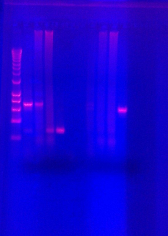
Step 3: Gel Electrophoresis
Once traditional PCR is complete, the sample will be fluorescently dyed and transferred to a gel which uses electricity to separate DNA molecules by size. Each gene targeted has a known size that if present will separate at an expected location on the gel as a band of light. Using UV light we can visualize the separated DNA bands to determine if the pathogen DNA is present.
Polymerase Chain Reaction is a highly accurate technique used in laboratories all over the world. This technology, when used correctly, can give very precise results of pathogen infection in ticks.
Contact Us
Contact Information
- Campus Address
- ESU Innovation Center
- Phone:
- (570) 422-7892
- Fax:
- (570) 422-3724 (Fax)
- Title of Department Leader
- Director
- Name
- Nicole Chinnici
- E:
- nchinnici@esu.edu
- Phone:
- (570) 422-7891